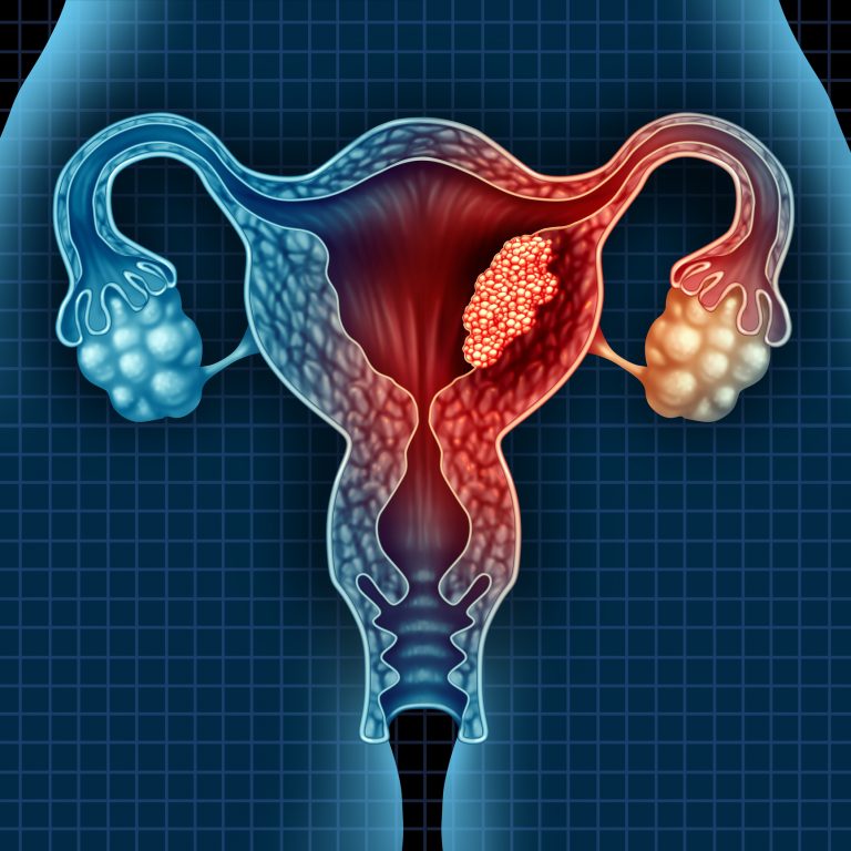
A study led by the University of Cambridge has completed an in-depth cell map of the uterus and endometrial tissue, leading to insights into the cellular basis of different forms of uterine cancer.
Published in the journal Nature Genetics, the study is the first in-depth reference map of the uterus to collect both single cell and spatial transcriptomic data.
The researchers identified two previously unknown epithelial cell states that can differentiate between different types of uterine cancer. The cell states SOX9+LGR5+ and SOX9+LGR5- increase in number as the endometrial cells regenerate during the menstrual cycle.
The team found the numbers of these cells present in the endometrium was linked with two different types of endometrial cancer. Tumors carrying a higher proportion of SOX9+LGR5+ were linked to more severe cases of cancer.
As many as one in three women will experience some form of reproductive disease during their life, including conditions such as endometriosis and life-threatening uterine cancers.
“There has been limited progress in the study of these disorders over the past decade, partly due to the challenges in analyzing this highly dynamic and complex tissue,” write the authors.
To produce the map the researchers analyzed womb tissue samples taken from 15 women of reproductive age using both single cell analysis and spatial transcriptomics, which uses mRNA analysis to map cells to their locations in a tissue sample. Then then used the information they gathered to build sophisticated endometrial organoids.
Overall, 98,568 cells were identified, which could be mapped to 14 cell types. There were five main cell categories in this group: immune, epithelial, endothelial, supporting, and stromal.
To find out more about which of the cell types are involved in uterine cancers, the team carried out a comparison of their map with endometrial cancer samples from the Cancer Genome Atlas.
They found SOX9+ epithelial cells were common in endometrial tumors. There were three main patterns of cells seen in endometrial adenocarcinomas. SOX9+LGR5+ was seen in 63% of serous and 24% of endometrioid adenocarcinomas; SOX9+LGR5−, was found in 33% of endometrioid adenocarcinomas but not in serous adenocarcinomas; and a small percentage (less than 10%) of endometrial cancers had expression of differentiated ciliated or glandular signals.
“Understanding the type and severity of a cancer can be crucial in deciding the best course of treatment,” said Roser Vento-Tormo, a senior author of the study from the Wellcome Sanger Institute.
“Our study shows that the combination of genomics, imaging and organoids can create a robust platform for studying endometrial physiology. This will have a wide-ranging impact on women’s health and reproductive medicine,” concludes the team.
The study was carried out as part of the Human Cell Atlas initiative, which is aiming to map every cell in the human body. Over 2,000 people across over 75 countries are involved in creating the Human Cell Atlas, and data are published openly and made accessible to researchers as the project is progressing.


